主题:True molecular resolution imaging: Fluorescence imaging with a spatial resolution in the one-digit nanometer range
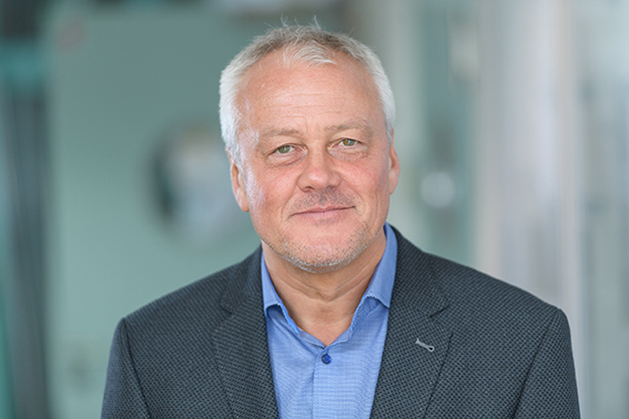
报告人:Markus Sauer,Julius Maximilian University Würzburg, Germany
时间:2024年3月18日(周一) 13:00-15:00
地点:霞光楼220会议室
邀请人:任吉存 教授

个人简介
Markus Sauer studied Chemistry at the University Heidelberg where he received his Diploma in 1991 and his PhD in 1995 in Physical Chemistry. 1998 he has been awarded the BioFuture Prize for Detection, Analysis and Handling of Single Molecules, which allowed him to establish his own group for single-molecule fluorescence detection and single-molecule DNA sequencing. From 2003-2009 he was Professor and chair of Laser Physics and Laser Spectroscopy at the University Bielefeld, Germany. Since 2009 he is Professor and Chair of the Department of Biotechnology and Biophysics at the Julius Maximilian University Würzburg, Germany. His research interests are single-molecule fluorescence spectroscopy and imaging with a particular focus on super-resolution fluorescence imaging by direct stochastic optical reconstruction microscopy (dSTORM) and its applications in neurobiology and immunology. He has published more than 300 journal papers and coordinates several super-resolution microscopy projects.
报告摘要
Super-resolution microscopy has evolved as a very powerful method for sub-diffraction resolution fluorescence imaging of cells and structural investigations of cellular organelles. Super-resolution microscopy methods can now provide a spatial resolution that is well below the diffraction limit of light microscopy, enabling invaluable insights into the spatial organization of proteins in biological samples with a spatial resolution of 20-30 nm. Recently, refined single-molecule localization microscopy methods such as MINFLUX and DNA-PAINT achieved superior localization precisions enabling the separation of two fluorophores separated by only 1 nm. However, translation of such high localization precisions into sub-10 nm spatial resolution in biological samples remains challenging. I will discuss different possibilities to bypass these limitations. One is based on physical expansion of the cellular structure by linking a protein of interest into a dense, cross-linked network of a swellable polyelectrolyte hydrogel. By combining Expansion Microscopy (ExM) with super-resolution Structured Illumination Microscopy it is possible to realize four-color fluorescence imaging in cells with ~20 nm spatial resolution. I will show how the spatial resolution can be further pushed to the one-digit nanometer range in cells by combining ExM with dSTORM. Another approach I will discuss in the use of resonance energy transfer between fluorophores separated by less than 10 nm. Using time-resolved fluorescence detection in combination with photoswitching fingerprint analysis interfluorophore distances of only a few nanometers can resolved. We will show how the method can be used advantageously to determine the number and distance even of spatially unresolvable fluorophores in the sub-10 nm range. I will demonstrate the performance of the different one-digit nanometer imaging methods using biological reference structures and protein-based PicoRulers generated by genetic code expansion (GCE) with unnatural amino acids and bioorthogonal click-labeling with small fluorophores. Finally, I will demonstrate that time-resolved photoswitching fingerprint analysis can pave the way for true molecular-resolution fluorescence imaging in fixed and living cells.


